Common Accessory Ossicles of the Foot UW Emergency Radiology
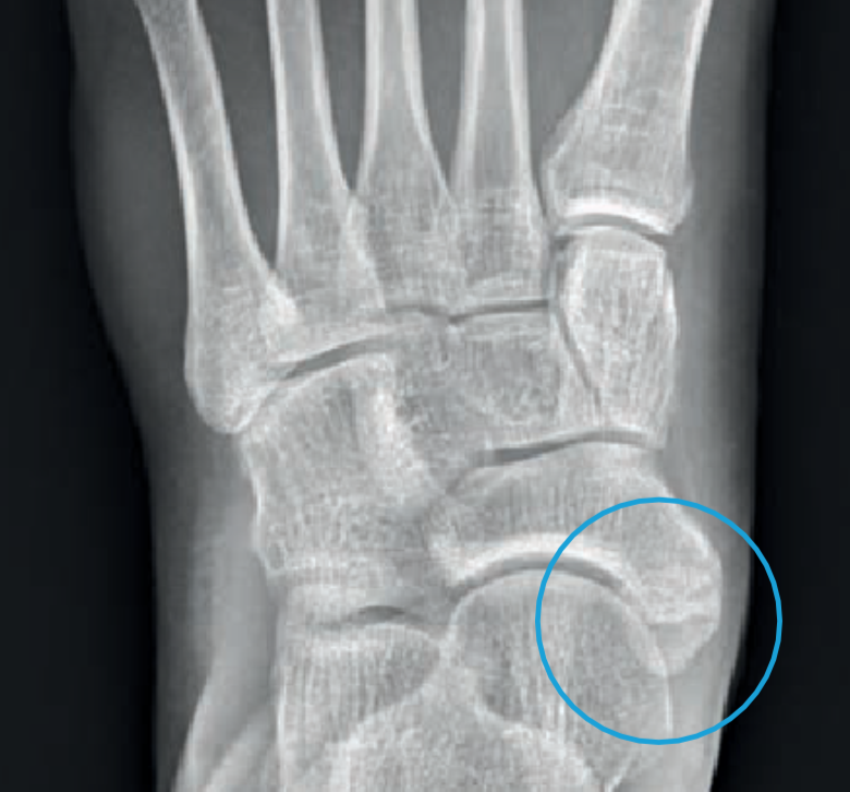
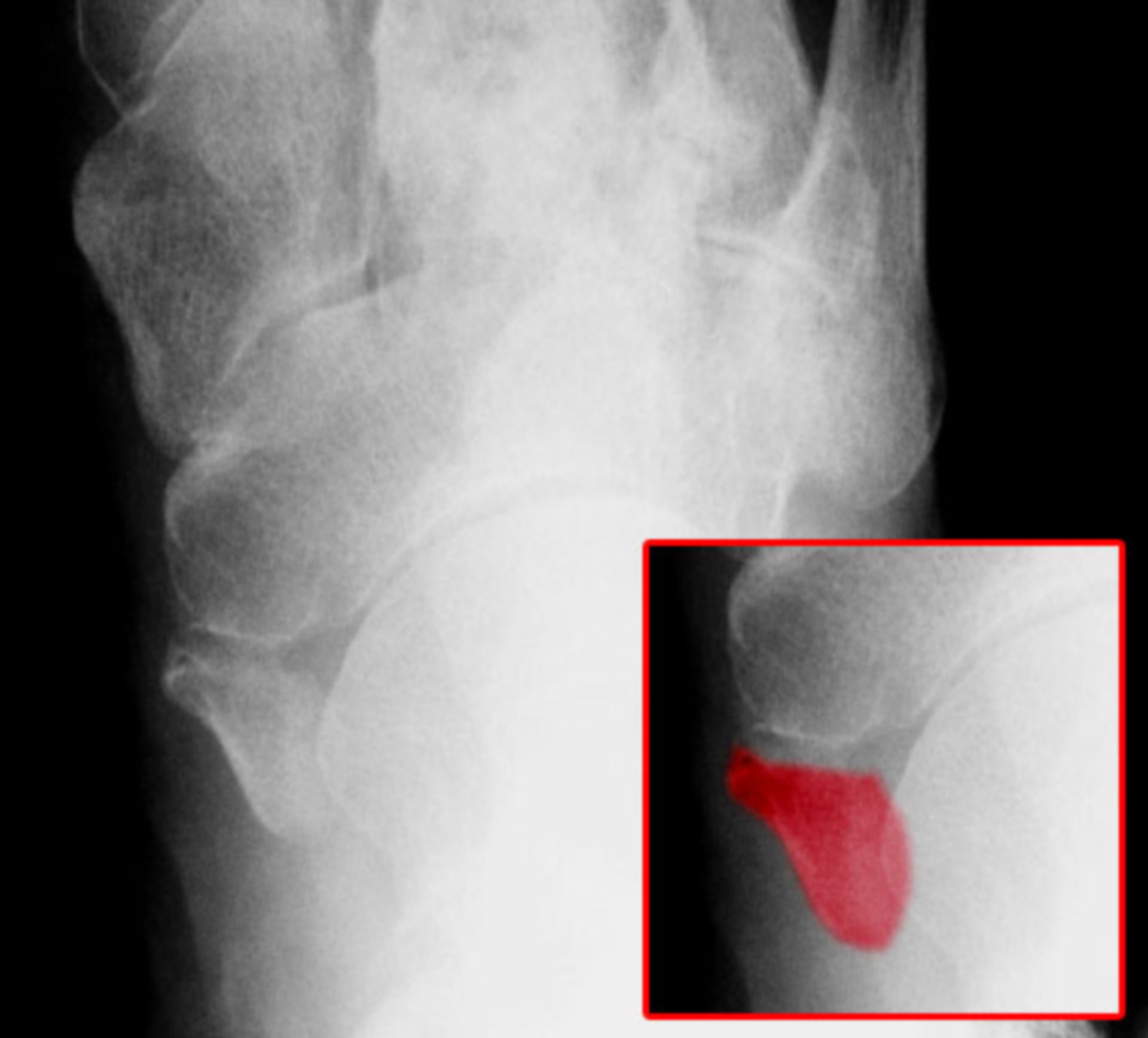
The accessory navicular—also known as the os naviculare or os tibiale externum—is a small bone that extends from the navicular bone, one of the tarsal bones near the instep. About 14 percent of the population has an accessory navicular, and about half of the people with the extra bone have it in both feet.
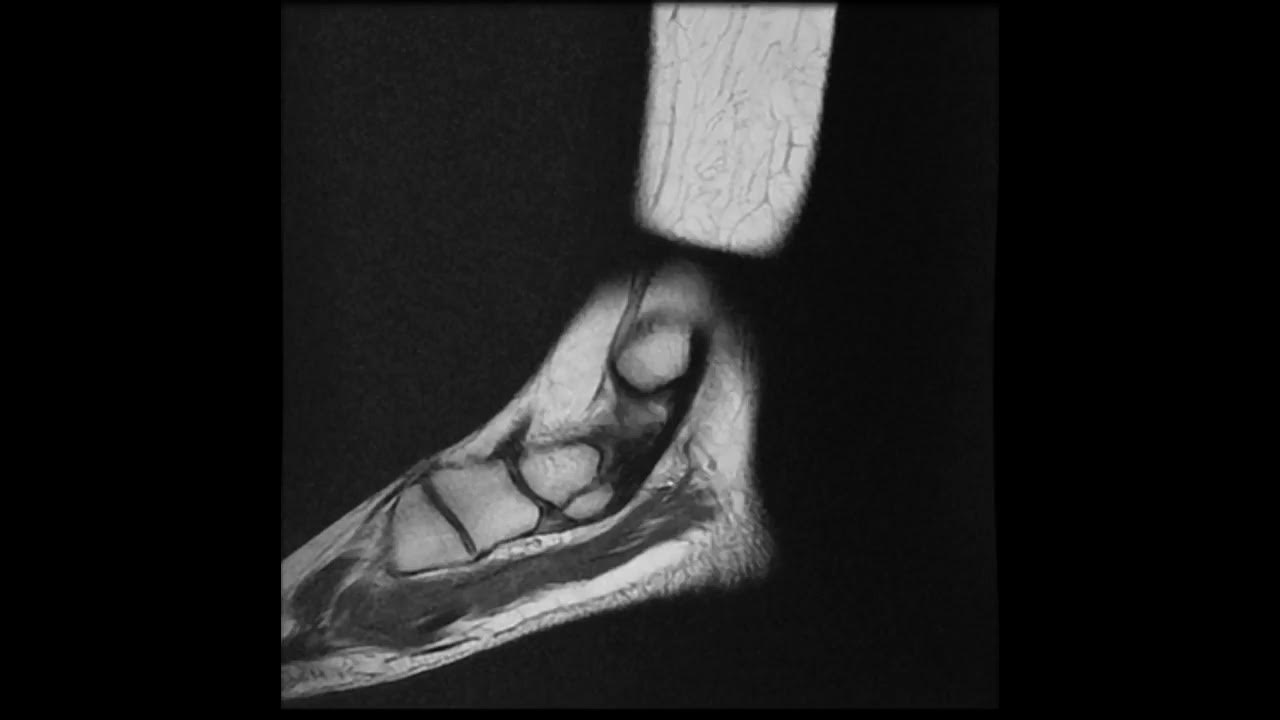
Os tibiale externum sagittal T2 YouTube
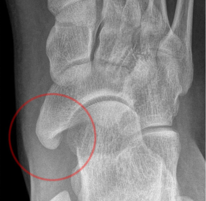
An accessory navicular is a large accessory ossicle that can be present adjacent to the medial side of the navicular bone. The tibialis posterior tendon often inserts with a broad attachment into the ossicle. Most cases are asymptomatic but in a small proportion, it may cause painful tendinosis due to traction between the ossicle and the navicular.

Surgery Assistant
An accessory navicular is a large accessory ossicle that can be present adjacent to the medial side of the navicular bone. The accessory navicular bone presents as a sesamoid in the posterior tibial tendon, in articulation with the navicular [1] or as an enlargement of the navicular itself. Epidemiology Navicular bone green

Lower Extremity Os Foot & Ankle Orthobullets
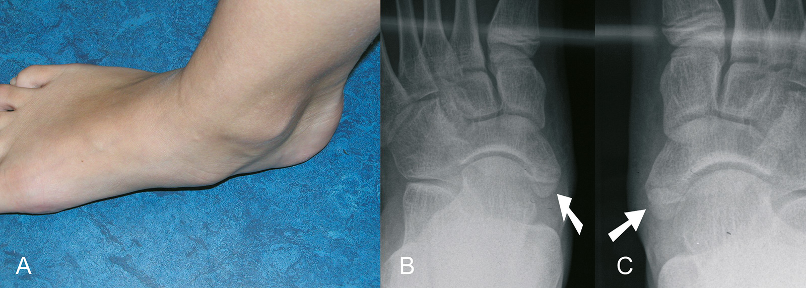
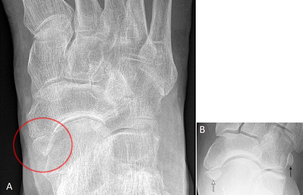
Acessory Navicular is a common idiopathic condition of the foot that presents with an enlargement of the navicular bone. Diagnosis is made with plain radiographs of the foot showing a plantar medial enlargement of the navicular bone.
Orthoforum Akzessorische Knochenkerne
The accessory navicular bone is a surplus piece of cartilage or bone fragment. It usually forms in the inner part of the foot, right above the arch. It's called the accessory navicular since it.

Os tibiale externum type II Image
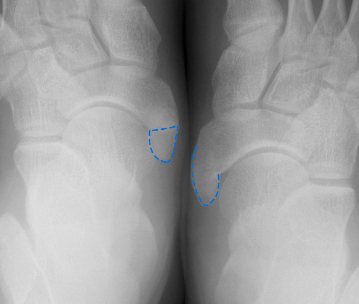
Type 1: An os tibiale externum is a 2-3 mm sesamoid bone in the distal posterior tibialis tendon. Usually asymptomatic. Type 2: Triangular or heart-shaped ossicle measuring up to 12 mm, which represents a secondary ossification center connected to the navicular tuberosity by a 1-2 mm layer of fibrocartilage or hyaline cartilage.

Beyond the obvious Exploring Os Tibiale Externum and Os Peroneum in Foot and Ankle Pain A
Accessory ossicles usually remain asymptomatic, but can become painful due to fractures, dislocations, degenerative changes, osteonecrosis, osteoarthritis, osteochondrial lesions, avascular necrosis, tumors, and irritation or impingement of adjacent soft tissues.
ostibialeexternumtypeiiandiii2 KENSHIN blog
Also known as os naviculare or os tibiale externum, an accessory navicular is an extra bone on the inside of the navicular (the bone in the middle of the arch of the foot) and within the posterior tibial tendon that attaches to the navicular bone. Top-view of accessory navicular in the right foot Types of accessory navicular

Os tibiale externum sagittal T2 YouTube
Characteristics and articulations The navicular is a boat-shaped bone, which has an important role in the maintenance of the medial longitundinal arch of the foot. Proximally, the navicular bone consists of a concave surface with an ovoid shape that articulates with the head of the talus.

Lower Extremity Os Foot & Ankle Orthobullets
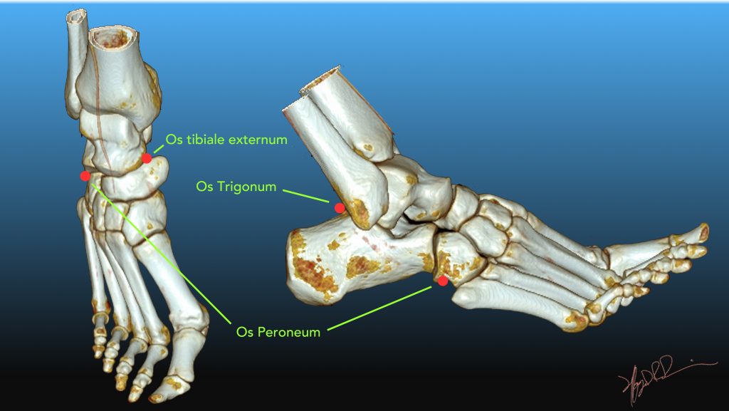
The os tibiale externum is also known as accessory navicular bone, os naviculare secundarium, accessory (tarsal) scaphoid, or prehallux. It is found within the tibialis posterior tendon near its insertion on the navicular bone. The os peroneum is a small sesamoid bone located within the peroneus longus tendon, adjacent to the cuboid.
Knickfuss Leonardo
also known as os tibiale externum 2-3 mm sesamoid bone embedded within the distal portion of the posterior tibial tendon no cartilaginous connection to the naviculam tuberosity and may be separated from it by up to 5 mm accounts for 30% of accessory navicular bones usually asymptomatic type 2 accessory navicular bone
Os tibiale externum DocCheck
Os tibiale externum (OTE) also termed accessory navicular, os naviculare, or os navicularis is a common accessory bone in the foot located medial and sometimes proximal to the navicular tuberosity. It is attached and continuous with the tibialis posterior tendon and is present in 10 to 15% of the population either unilateral or bilateral.
Os Tibiale Externum Ortobas
Definition accessory ossicles secondary ossification centers that remain separated from the normal bon sesamoids are bones that are incorporated into tendons and move with normal and abnormal tendon motion Most common ossicles os trigonum accessory navicular (os tibiale externum) os intermetatarseum Most common sesamoids os peroneum

Os tibiale externum Image
The accessory navicular syndrome , also known as os naviculare syndrome occurs when a type II accessory navicular becomes painful due to movement across the pseudo-joint between the ossicle and the navicular bone. Radiographic features Ultrasound
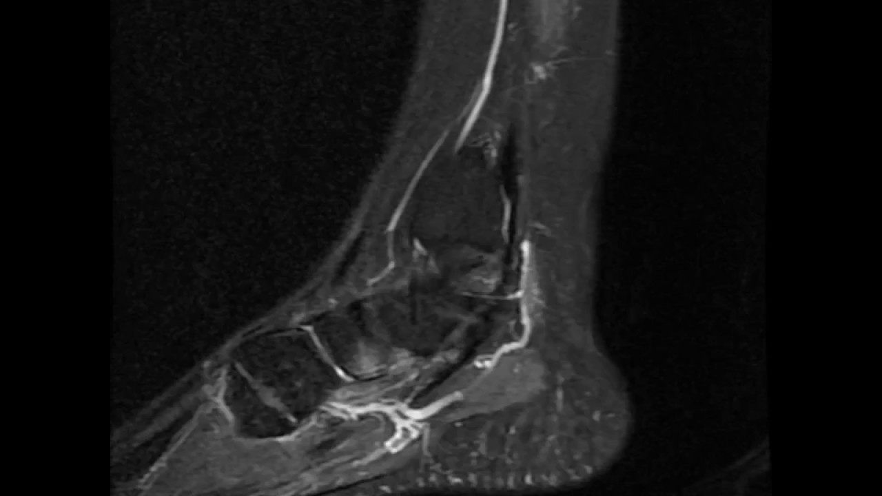
Os tibiale externum sagittal T2 fat sat YouTube
The accessory navicular (os navicularum or os tibiale externum) is an extra bone or piece of cartilage located on the inner side of the foot just above the arch. It is incorporated within the posterior tibial tendon, which attaches in this area and can lead to Accessory Navicular Syndrome. An accessory navicular is congenital (present at birth).
Os Tibiale Externum Ortobas
In rare cases, the accessory navicular bone creates a bony prominence in the midfoot that causes pain, redness and swelling in the medial arch area, plantar fasciitis, bunions and heel spurs. When this happens, the condition is called accessory navicular syndrome. ANS can arise from a number of things, including foot trauma like ankle sprains.